Foot Lameness

Foot Lameness
Lameness in the forefoot is by far the most common unsoundness in the horse. There are a multitude of reasons for this such as anatomy, shoeing and trimming methods and athletic activities. When one considers how small the horse‘s foot is relative to the body mass it is supporting just at rest, then add in the various forces that affect the foot with exercise it‘s remarkable that there are not more problems.
Anatomically the foot is a complex structure with bones that need to be in correct alignment. There are tendons and ligaments that insert into these bones and exert various forces depending on what action is being taken. Even at rest there are areas of tension. There are joint capsules and joint surfaces that are potential sites for inflammation when problems develop. Bursae, fluid filled structures that prevent irritation to a tendon as it passes over a bone, are located throughout the body. There is one bursa in each foot along the back edge of the navicular bone. As with joints these bursae can become inflamed. Other non-joint related tissues that can account for foot pain are the laminae or interconnected "fingers" that are partially responsible for maintaining the coffin bone in position along the hoof wall. A foot lameness may involve more than one of these structures at a single time.
Anatomy of the hoof:

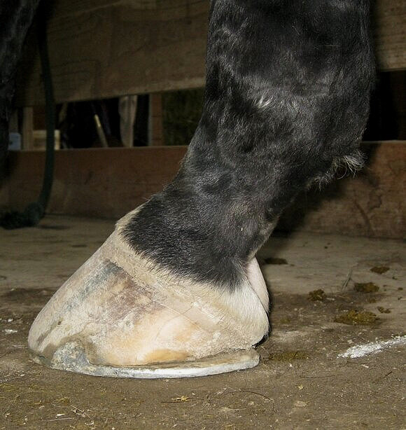
The underside of healthy foot with normal structures.

Evaluating the foot and internal structures from the side:
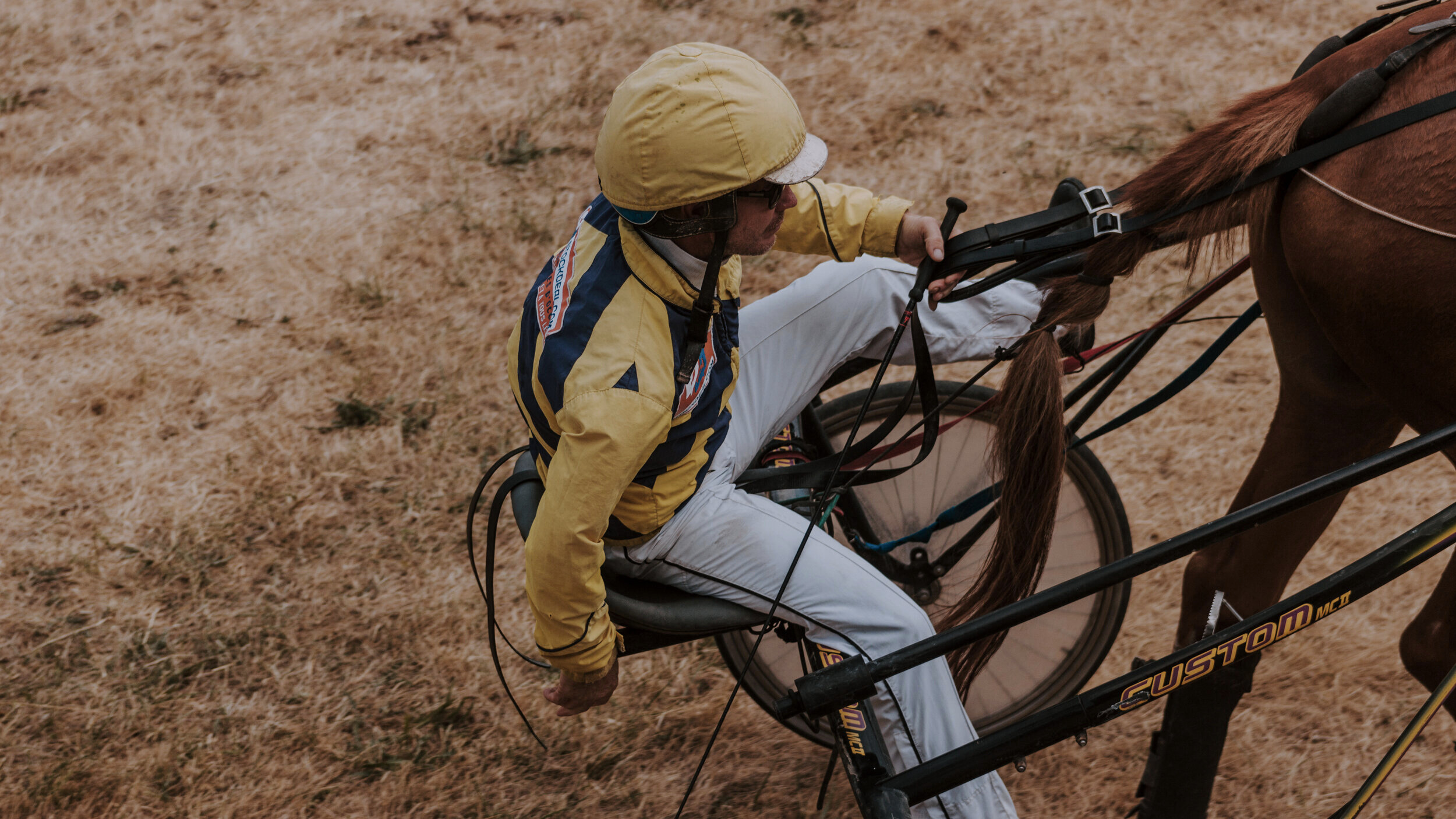
A low-heel foot. This horse is shod with a shoe set back from the toe and extended back to the heel bulbs to increase palmar support.

In the area just from below the fetlock down to the tip of the toe there are numerous structures that can each cause a lameness to develop. When they are combined and hidden by the hoof wall they may even be harder to diagnose.
Diagnostics Procedures
Diagnosing a lameness to the foot is not generally difficult, but localizing the source is the challenge. There are certain hallmarks for various lameness conditions that help to key us into particular areas. The first may be how the horse stands. Does the horse point the foot, prefer to keep the fetlock flexed and the foot in a non-weight bearing position or does the horse prefer to place the foot laterally? These positional changes generally indicate problems with the feet and pointing often indicates the presence of heel pain. Feeling for heat or an increase in pulses aids in identifying foot inflammation. The pulses may be easiest felt along the back of the fetlock and a comparison can be made with the opposite leg. A normal pulse may be barely perceptible to most people while one associated with foot inflammation may be markedly elevated. Another technique used to locate foot pain is the application of hoof testers. These tools are designed to apply pressure in selected areas such as the frog or sole. This may assist in diagnosing a sole bruise, an abscess, a close nail, thrush, founder and heel pain. If the problem presents itself immediately with hoof testers there may not be a need for a complete lameness exam. Each lameness tends to be somewhat unique and frequently does not allow for a routine diagnosis or treatment. If an abscess or bruise can be opened and treated then the recovery time is markedly shortened. There are times when an abscess may be deeper requiring re-evaluation after a period of soaking and poulticing. Initially a bruise may affect the foot in much the same way as an abscess and lead to questions on whether to pare out the sole further in search of an abscess or whether to pull the shoe. Bruises may take time to resolve, but they do not intensify like abscesses as pressure increases.
An example of a foot with 3 potential causes of a foot lameness:

Lateral x-ray of a forefoot with multiple issues. The chronic problems would be evidence of Ringbone (a degenerative disease of the pastern and coffin joints) and a large bone fragment in the dorsal aspect of the coffin joint. His more immediate issues involved sinking in his forefeet- a type of founder where the coffin bone displaces towards the ground surface.
Diagnostics
Diagnostic blocking is a procedure where a local anesthetic — a drug similar to Novocaine — is placed adjacent to nerves to selectively block or numb certain parts of the foot and leg. Once an area is blocked and the horse becomes sound we then know the region that is involved in causing a lameness. Joint blocks are done in a similar fashion except the procedure is more specific since the medication goes directly into a joint. Unfortunately there are no hard and fast rules with these diagnostic procedures. There can be inconsistencies with nerve supply and diffusion of blocking drugs thereby confusing attempts to make a clear diagnosis. Regardless of these problems localizing the source of the pain to a certain region still allows for more specific diagnostic procedures and focused treatment plans.
X-rays of the foot is a common procedure done on almost a daily basis. Attempting to visualize a fracture, evidence of founder, navicular degeneration, coffin bone inflammation (pedal osteitis) and/or degenerative joint disease (arthritis) is straightforward and best done through radiography. There are many soft tissue structures that cannot be seen on x-rays such as tendons, ligaments, joint capsules and bursae. Therefore not seeing a problem on foot x-rays does not necessarily rule out a foot problem it merely makes the diagnosis more elusive without using other diagnostic techniques.
Other techniques that provide information about the bones and soft tissues of this region are: ultrasound, thermography, nuclear bone scans, MRI and CAT scans. Obviously some of these procedures are not offered in the field, but they are becoming more available through veterinary schools and referral ceenters. Ultrasound is used frequently in the field and involves the use of sound waves to aid in visualizing various anatomical structures. Depending on where we are looking in the foot, ultrasound has limitations due to the proximity of the bones to the skin surface, which affect the echoes of the sound waves. Mainly we look at tendons and ligaments along the back of the pastern and at ligaments along joint edges. Tearing or enlargement of tendons or ligaments can easily be visualized and measured by ultrasound in the field. Thermography is a technique that uses heat emissions to locate a source of inflammation. Using a camera designed to detect gradations in heat output we are able to locate the presence of inflamed tissue. Another benefit of this procedure is looking at hoof balance. If the foot is landing unevenly heat will be generated based on the foot strike on the ground. As with any lameness, balancing the foot with respect to toe-to-heel and side-to-side ground contact is integral to decreasing the stress on hoof capsules, bones, ligaments and joints. We use thermography in the field and analyze the data both there and back at the office where we can generate pictures from the computer. See article on thermography. Nuclear bone scans are procedures done at referral centers such as Myhre Equine Clinic, New England Equine and Tufts University Veterinary School. The horses are given an intravenous injection of a drug with a radioactive agent. The drug circulates throughout the body and accumulates in areas with the most blood flow which will correspond to sites of inflammation. After an appropriate period of time a camera is used to detect the areas that are concentrating the radioactive drug thereby directing us to these sites of inflammation. Following this, x-rays, ultrasound and/or MRI can be done for more specific information. This procedure can be especially helpful when there are multiple sites, when the back or hips are possibly involved, when a consistent lameness is not always present and when traditional diagnostics are not successful or cannot be safely done. There are no risks to the horse with this procedure, the only downside is the cost (+/- $1,000=$1500), but when a diagnosis is elusive and response to therapy is limited, this can greatly enhance our ability to find and treat the problem more effectively.
The use of MRI and Cat or CT scans is limited to only a few facilities. The image detail is phenomenal, but the cost and limited availability preclude its use on a regular basis. It is best used on the extremities and provides detailed information about joint surfaces, ligament attachments around joints, tendon and ligament fibers and other soft tissue structures. It’s fortunate if a horse is insured as these procedures can be in the range of $2500 when done using general anesthesia, if done standing the costs may be closer to $1650.
Treatments
The therapies used in treating a foot lameness depend on the location of the problem and the nature of the condition. Often we are left with a regional diagnosis such as "caudal heel lameness". This identifies the heel area as the region, but leaves open the question of which if any soft tissues are involved with a "navicular type lameness". Therefore a combination of therapies is often the most appropriate route to undertake. In my opinion, balancing the foot with respect to proper trimming and shoeing is critical for any treatment to be successful. If the foot is not balanced — not landing evenly — and if the heel is too low and the toe too long thereby lowering the hoof angle, problems will persist. Stress relief in the foot is essential. The most important concepts are decreasing lateral to medial (side-to-side) instability, decreasing length of toe to ease breakover and providing adequate heel support. We must decrease the inertia at the toe as the foot rolls over and likewise decrease the tendon tension on the heel area as the heel is lifted and the foot is brought forward. Less resistance at the toe translates into less potential hoof wall stress and heel pain.
Besides trimming changes, farriers may use extended heel shoes, natural balance shoes, bar shoes and/or degree pads to accomplish the goal of improving heel support and possibly raising the hoof angle. Some horses require "therapeutic shoeing" for several shoeing cycles then return to a normal keg shoe once the heel grows out, while others may do best with a bar shoe and degree pad. See article on foot balance.
The use of medications in the treatment of foot pain is integral to a successful outcome and to improve the patient‘s overall comfort. Which medications and their duration of use are determined by the intensity, chronicity and response to initial therapy. Anti-inflammatories are drugs used to decrease pain and inflammation. The most commonly used is Bute (Phenylbutazone) since it consistently has the best results with musculoskeletal pain. Initially it may be used at a higher dose then tapered off over a week or two. In the case of an older horse with chronic problems and the likelihood of multiple joint involvement, this may be used on a daily basis. Generally our plan is to decrease the anti-inflammatories while foot balance is restored and foot conformation improves. Bute is considered a Nonsteroidal Anti-Inflammatory Drug (NSAID), in addition the following would also be members of the same group: Previcox, Equioxx, Banamine and Ibuprofen. Other drugs used in a heel lameness are those that alter blood flow to the area. Their use is somewhat controversial as some Veterinarians feel there is minimal empirical evidence that justifies their use and furthermore a blood flow problem may not be that important in a heel lameness. In veterinary equine practices drugs that affect circulation — Isoxsuprine and Pentoxyfilline — may be used routinely and play an integral role during the treatment phase and in some instances as a maintenance drug. There are some horses that remain on Isoxsuprine only and relapse when the dose is decreased.
Intra-joint treatments are used if the initial therapies have limited benefit, if we decide to be more aggressive from the start or if we see signs of degenerative joint disease in the coffin joint. The medications are usually a combination of a steroid and hyaluronic acid — the normal joint fluid component. The steroid or cortisone will rapidly decrease joint inflammation and the hyaluronic acid will help with lubrication of joint edges and assist in rebuilding joint fluid. In addition to coffin joint injections, cortisone injections into the navicular bursa are being used very effectively to treat some of these horses. In fact for some of these "caudal heel lameness" cases that have had feet balanced and been through an appropriate period of rest so the use of anti-inflammatories are probably the ones best suited for the bursal injection.
Newer medications that fall into the family of drugs called Bisphosphonates may also play an important role when treating bone edema and altered bone responses to inflammation. Frequently Osphos is used for these cases, in earlier years we used Tildren more frequently - their mode of action is the same, they differ in how they’re administered. Research has demonstrated some remarkably effective responses with horses treated with these medications and having a caudal heel (navicular type) lameness plus bone degeneration also ones with Kissing Spine Disease.
Shockwave Therapy (SWT) is another treatment that is being offered for caudal heel lameness at Burlington Equine. So far the results have been mixed. There are cases with dramatic improvement then others where limited benefits are appreciated. Between our ability to better image the affected area with digital x-rays, U/S and advanced modalities then treat with a variety of options, such as injections and wave therapy, the diagnostics and treatments have been taken to a new level. In fact, we have seen some or the chronic heel lame horses respond very well to one or two shockwave sessions, where we were having limited results to medications. As MRI becomes more available at referral centers this may be included as a diagnostic option and focusing the treatments on other causes may be pursued.
Laser Therapy, another modality that uses waves and, in this situation ones generated with light energy, has the ability like SWT to promote healing in the traumatized tissue, improve blood flow and reduce inflammation. The Class IV laser used by Burlington Equine has excellent penetration of the affected area and an ability to disperse the wave energy more diffusely throughout the region than we see with SWT. In an instance where there is an interface between bones and ligaments the SWT is considered the preferred approach, but many of the laser and SWT procedures overlap.
There are systemic joint medications designed to treat generalized joint inflammation and to help maintain soundness. These drugs — Adequan and Legend — have helped horses with foot pain either as an adjunctive therapy or as the only drug. They have limited benefit if the problem involves tendons and ligaments plus hoof balance. Their purpose is to improve the quality of the joint fluid, protect the cartilage from further degradation and to decrease joint inflammation. If the lameness involves joint structures such as the coffin joint or navicular bursa then their use is indicated. There are absolutely no negative side effects and in terms of promoting overall joint health their effectiveness is unparalleled.
As previously suggested we will often initiate treatment with a combination of approaches. Improvement may take as little as 2 weeks, but some can stretch to 4-6 weeks or even up to 6 months+ before an effective treatment response is realized. Those that take longer may require more rest and time off from training. A slow return to work while allowing previously injured tissues to undergo some stress without triggering the pain or inflammatory cycle is important. Physical therapy is a concept much better understood with human injuries than in those involving horses.
Surgery may be the final option for horses not responding to other treatments. A "nerving surgery" for a caudal heel lameness is not nearly as commonplace as it was years ago due to better treatment strategies and diagnostic techniques. However, there are some horses that are not going to respond to any other therapies. Nerving involves severing the two nerves along the back of the pastern that supply sensation to the heel area. The horse will still be able to feel the toe, but heel pain and sensation will be removed. This tends to ensure a semi-permanent solution to the problem. The reason that it may not be permanent is due to the re-establishment of nerve supply over time. The surgical technique and experience of the surgeon often are the critical factors in determining the best outcome. Understandably there are risks associated with this procedure, as with all surgeries, and these must be weighed against the potential benefits. Every year we refer several horses that are recalcitrant to other medical therapies and find that the outcome is very satisfactory. In fact, there are upper level dressage and jumping horses that would be unable to compete without this surgery.
Diagnosing a foot lameness is relatively straightforward, yet identifying affected structures and tissues with reliable specificity remains the challenge. Fortunately treatment strategies have improved and when necessary diagnostic procedures exist which can bring out an elusive diagnosis. Most of these lamenesses, even those originating from the navicular region, do not have the poor prognosis as was commonly thought. Perseverance in treatments will be the key to ensuring success.