Digital Radiographs

Digital X-rays
Digital x-rays have become the standard for equine practice and they have been used in this practice for the past 15 years. The amount of detail, ability to focus and manipulate an image and the ease of storing and transmitting x-rays electronically have made them superior than the xrays on film that most of us were trained with. Being able to diagnose conditions in the field and to work with farriers and to consult with surgeons within minutes of taking an image allows for a more accurate diagnosis and effective therapeutic plan. For those of you not familiar with this technology, simply think about the benefits of digital photographs or sound. Digital x-rays allow us to take an image in the field and have it displayed on a computer screen within seconds. Images can then be expanded, altered to enhance different aspects of the tissue and analyzed quickly to provide a diagnosis or to complete a purchase exam. Although traditional x-rays can still be beneficial, the digital x-rays offer unsurpassed image quality and the ability to diagnose conditions that could remain elusive even with our best techniques.

Once images are taken they can be saved onto an external drive for longer term storage, uploaded to a server off site and/or sent to another person electronically or shipped on a disk or flash drive. In addition images can be saved as jpeg files for clients to show farriers to aid with a shoeing plan. See article on Hoof Balance and Farrier Consultations
Digital radiography has rapidly developed in equine medicine and now with battery powering plus Bluetooth the images can be more easily obtained.
Examples of Digital Radiographs
Below are examples of regions frequently radiographed other areas are also routinely imaged.
Back Radiographs:
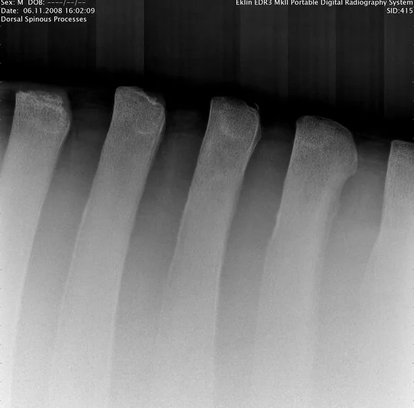
The vertical portion of the vertebrae should be evenly spaced as is seen with this normal radiograph of a horse's spine. Compare this with the one to the right that was taken in the saddle region.

As you will see in this radiograph versus the one to the left, the vertebrae are touching or rubbing against one another. This x-ray was taken without the weight of a rider. Once there is weight on the horse's back the problem would be significantly worsened. This is called "Kissing Spine" syndrome.
Stifle Radiographs:


Smooth joint surfaces and adequate spacing between joints. Ultrasound would be required to investigate soft tissue structures.
Neck Radiographs:


Skull Radiograph:

Foot Radiographs:
Since we have a strong interest in working with farriers we expect to be able to supply high quality images that will permit measurements and an ability to effectively monitor various foot conditions.


Article discussing foot balance and farrier consultations.
Digital radiography has rapidly developed in equine medicine and now with battery powering plus Bluetooth the images can be more easily obtained.
